Lateral Inhibition in Pink
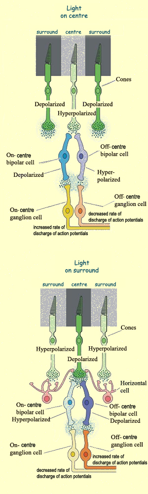
The optical illusion depicted illustrates an example of lateral
inhibition. Lateral inhibition promotes increased contrast sensitivity
through the detection of borders, as enhanced by feedback modulation of
signals by horizontal cells. Horizontal cells come in three types,
depending on the types of photoreceptors they synapse with, but that
detail is less relevant to their function in lateral inhibition overall.
Light detection is performed by the photoreceptors, which hyperpolarize
in the presence of light and depolarize in the absence of light (an
exception to most neuronal signaling). Depolarization leads to increased
glutamate release, the neurotransmitter currency of photoreceptors,
while hyperpolarization leads to decreased glutamate release. Whether
the receptive field is on-center or off-center is relevant to the types
of bipolar cells downstream, but is only really relevant to this
discussion in the sense that the center and surround provide contrast to
one another. The excitation or inhibition affects two types of bipolar
cells (on-center and off-center), which then affect their corresponding
retinal ganglion cells (also on-center and off-center). These cells
downstream of the photoreceptors and horizontal cells are interesting in
themselves, but not necessary for a discussion of lateral inhibition,
which is mostly about contrast. Horizontal cells receive excitatory
input from photoreceptors and send inhibitory input to all/neigboring
photoreceptors. In this case (of an off-center receptive field), the
surrounding horizontal cells are hyperpolarized in the presence of
light. When horizontal cells are hyperpolarized, they send less
inhibitory signals (recall that they send only inhibitory signals). The
neighboring photoreceptor in an off/dark region is already depolarized
in the absence of light, but because it receives less inhibition from
its neighboring horizontal cells, it depolarizes even more and releases
even more glutamate. It is this "even more" effect of modulation by
horizontal cells that increases border contrast and causes shades to
stand out more when placed next to one another.
Mental and Visual Distress during Pandemic Times
In a second study about Charles Bonnet Syndrome published this year,
researchers at University College London seek to highlight the increased
mental distress during the COVID-19 pandemic for those suffering from
the condition. The study itself is small, comprising only 45 patients at
one hospital. Among these patients, the researchers found that 56%
experienced increased intensity and exacerbation of visual
hallucinations during the interval of study between June
and July 2020. The weight of the study lies in its social focus to call
attention to Charles Bonnet Syndrome. The visual hallucination symptoms
of this condition are frightening in themselves in ordinary times. And
mental distress is unfortunately common for many during the pandemic.
That the mental distress of "social
isolation, loneliness, lack of exercise and exposure to distressing
media over the COVID-19 lockdown" increased the symptoms of Charles
Bonnet Syndrome highlights the need for awareness of the condition in
particular, but also the need for social and community support for the majority of people suffering from isolation and loneliness during the pandemic.
Disulfide Bridge Formation in the Crystalline Lens
Researchers at Goethe University in Germany are taking a basic science
approach to study the protein structure of the crystalline lens using a
genetically modified bacterial model. In particular, they studied the
disulfide bridges, bonds between two sulfur-containing amino acids, in
βγ-crystallins. Where this is interesting from a molecular standpoint is
that disulfide bonds are not easy for the cell to produce and maintain
given a cellular environment that promotes their dissolution. In the
finished βγ-crystallin protein, these bonds are shielded from the
cellular environment by placement in the internal parts of the protein.
But while the protein is in the process of formation, this is not yet
possible. It had been previously assumed that because the ribosomal
tunnel is too narrow, disulfide bridges could only form after protein
formation was completed. The researchers tested this assumption on their
bacterial model using various spectroscopic techniques and theoretical
simulations. They found that disulfide bridges are already formed during
the synthesis of the amino acid chain, that is, within the ribosomal
tunnel. One of the researchers notes, "Surprisingly, however, these are
not the same disulphide bridges that
are later present in the finished gamma-B crystallin. We conclude that
at least some of the disulphide bridges are later dissolved again and
linked differently," and that perhaps the preliminary disulfide bridges
accelerate the formation of the final disulfide bridges when the protein
is released from the ribosome. The focus and weight of this study in
the basic sciences, and in a very restricted aspect of crystalline
protein formation, limit its application in clinical settings.
Nonetheless, the crystalline lens is a structure with ubiquitous impact,
the transparency and power of the eye being due in large part to the
packing of these crystallin proteins in the lens (with the nuance that
the major refractive component of the eye is the cornea). Study of
crystalline lens formation thus informs the progression of cataracts and
could aid in research toward their prevention.
Protein Imbalance in Congenital Cataracts in Mice
This is another small study from Germany about the crystalline lens and
cataract formation. Researchers at the Technical University of Munich
used a mouse model to study hereditary forms of cataracts. The findings
were not major, however, the researchers discovered that at least for
the genetic conditions under study in their mouse models, clumping of
normal rather than abnormal proteins was the cause of cataracts. The
unstable, mutated proteins were eliminated immediately and not present.
Instead, it was the "healthy" normal proteins that clumped together. One
of the researchers states that the model showed that "the balance
between the various proteins, or their ratios to one
another, is important. When one of these components is missing, the
remaining ones interact and form clumps." The proportion of hereditary,
compared to age-related or other acquired, cataracts is extremely small,
which limits the scope of the study. Nonetheless, as with previous
studies about the crystalline lens, this study adds to the basic science
knowledge base that could aid in developing drugs that help prevent or
delay cataract formation. Perhaps also in a philosophical way, the study
highlights the body's balance of components in maintaining optimal
function.
In the News: Edition 2
As with the first EyeNet opinion piece of 2021, I generally agree
with this perspective, this time about genuine representation, about
publishing integrity, about the invisible hard work of editors. I agree
with it more than meets the eye. Where this opinion piece is relevant to
eye care is in its reference to the use of hydroxychloroquine for the
treatment of COVID-19. Eye doctors are familiar with the drug by way of
monitoring for adverse effects of retinal toxicity. Hydroxychloroquine
(Plaquenil) was originally a drug for malaria, and also has uses in
various rheumatologic and dermatologic conditions. Used for these
inflammatory and skin-related conditions, hydroxychloroquine is an
effective drug. One might even say that the drug design was catered for a
specific disease or range of diseases. Because of hydroxychloroquine's
popularity, some in the medical community sought its use as a potential
treatment for the novel coronavirus. Some even imposed upon this drug
the unintended use in pediatrics,
which demonstrates the strength of the desire for a cure against
COVID-19. Hydroxychloroquine is not FDA-approved for this form of
treatment—a fact often repeated, to say the least—though
the scientific curiosity and humanistic motivation to pursue this line
of therapy were commendable. That being said, transparency of the
scientific process was a prudent move on the part of researchers, so as
to avoid false advertisement and misrepresentation while testing this
therapy. That hydroxychloroquine works exceedingly well for some
conditions does not mean it should be coaxed into use for other
conditions. Despite hydroxychloroquine's abysmal results as a treatment
for COVID-19, we can both—nay, we can all—laugh at the fact that it still appears in the news.
In Everyday News
(1) Ways to avoid COVID lockdown eye strain
(2) Free infant vision screening program
(3) The art of vision: how pets see the world
(4) Hairy corneal dermoid in a deer
Saturday, February 27, 2021
Week in Review: Number 6
Friday, February 26, 2021
Protein Imbalance in Congenital Cataracts in Mice
Article: Cloudy eyes caused by protein imbalance
Source: Technical University of Munich (Germany), via ScienceDaily
Published: February 16, 2021
 |
| Cataract-associated crystallin mutants are thermodynamically destabilized |
My rating of this study: ⭐
Further reading: Understanding the molecular mechanisms underlying cataract development
Schmid PWN, Lim NCH, Peters C, et al. "Imbalances in the eye lens proteome are linked to cataract formation." Nature Structural & Molecular Biology. 28:143-151. 11 January 2021. https://doi.org/10.1038/s41594-020-00543-9
Thursday, February 25, 2021
Mental and Visual Distress during Pandemic Times
Article: Visual hallucinations among blind people increase during pandemic
Source: University College London (U.K.), via Technology Networks
Published: February 12, 2021
In a second study about Charles Bonnet Syndrome published this year, researchers at University College London seek to highlight the increased mental distress during the COVID-19 pandemic for those suffering from the condition. The study itself is small, comprising only 45 patients at one hospital. Among these patients, the researchers found that 56% experienced increased intensity and exacerbation of visual hallucinations during the interval of study between June and July 2020. The weight of the study lies in its social focus to call attention to Charles Bonnet Syndrome. The visual hallucination symptoms of this condition are frightening in themselves in ordinary times. And mental distress is unfortunately common for many during the pandemic. That the mental distress of "social isolation, loneliness, lack of exercise and exposure to distressing media over the COVID-19 lockdown" increased the symptoms of Charles Bonnet Syndrome highlights the need for awareness of the condition in particular, but also the need for social and community support for the majority of people suffering from isolation and loneliness during the pandemic.
 |
| Response distributions for survey items among 45 people with Charles Bonnet syndrome |
My rating of this study: ⭐🌸
See also: A Brain Mechanism underlying “Vision” in the Blind is Revealed
Jones L, Ditzel-Finn L, Potts J, et al. "Exacerbation of visual hallucinations in Charles Bonnet syndrome due to the social implications of COVID-19." BMJ Open Ophthalmology. 6:. 11 February 2021. https://doi.org/10.1136/bmjophth-2020-000670
Wednesday, February 24, 2021
Disulfide Bridge Formation in the Crystalline Lens
Article: How the 3-D structure of eye-lens proteins is formed
Source: Goethe University (Germany), via ScienceDaily and Technology Networks
Published: February 10, 2021
 |
| Side view of a ribosomal 50S subunit with a focus on the nascent chain |
My rating of this study: ⭐⭐
Schulte L, Mao J, Reitz J, et al. "Cysteine oxidation and disulfide formation in the ribosomal exit tunnel." Nature Communications. 11(5569). 4 November 2020. https://doi.org/10.1038/s41467-020-19372-x
Tuesday, February 23, 2021
Lateral Inhibition in Pink
Article: How Many Colors in This Image? Here's The Science Behind The Illusion...
Source: ScienceAlert
Published: February 10, 2021

 |
| Center-surround receptive field, including interaction between photoreceptors and horizonal cells |
My rating of this article: ⭐⭐
Further reading: Center–Surround Receptive Field
Monday, February 22, 2021
In the News: Edition 2
Article: Of Fake Scholarship and Academic Integrity
Source: EyeNet
Published: February 20, 2021
As with the first EyeNet opinion piece of 2021, I generally agree with this perspective, this time about genuine representation, about publishing integrity, about the invisible hard work of editors. I agree with it more than meets the eye. Where this opinion piece is relevant to eye care is in its reference to the use of hydroxychloroquine for the treatment of COVID-19. Eye doctors are familiar with the drug by way of monitoring for adverse effects of retinal toxicity. Hydroxychloroquine (Plaquenil) was originally a drug for malaria, and also has uses in various rheumatologic and dermatologic conditions. Used for these inflammatory and skin-related conditions, hydroxychloroquine is an effective drug. One might even say that the drug design was catered for a specific disease or range of diseases. Because of hydroxychloroquine's popularity, some in the medical community sought its use as a potential treatment for the novel coronavirus. Some even imposed upon this drug the unintended use in pediatrics, which demonstrates the strength of the desire for a cure against COVID-19. Hydroxychloroquine is not FDA-approved for this form of treatment—a fact often repeated, to say the least—though the scientific curiosity and humanistic motivation to pursue this line of therapy were commendable. That being said, transparency of the scientific process was a prudent move on the part of researchers, so as to avoid false advertisement and misrepresentation while testing this therapy. That hydroxychloroquine works exceedingly well for some conditions does not mean it should be coaxed into use for other conditions. Despite hydroxychloroquine's abysmal results as a treatment for COVID-19, we can both—nay, we can all—laugh at the fact that it still appears in the news.