A Group of Cells in the Brain's Temporal Pole Specializes in Recognizing Familiar Faces
The fabled "grandmother neuron," a neuron that in itself encodes the
recognition of an individual person—such as one's grandmother or a
celebrity—is a theoretical concept at the culmination of visual
processing from the retina to the brain, including input from memory and
emotions. While such a singular neuron remains hypothetical,
researchers have discovered a group of "hybrid" neurons in the brain’s
temporal pole (TP) region that represents the connection between the
sensory and memory domains. These neurons behave like sensory cells in
the sense that they have reliable and fast responses to visual stimuli,
while at the same time responding only to stimuli that the brain has
seen previously, much like memory cells. Earlier research by the
investigators localized a small region in the anterior temporal cortex
of the brain as involved in facial recognition. In the new experiments,
they explored this region with fMRI in two Rhesus macaques as the
monkeys watched familiar and unfamiliar faces. The researchers found
that the neurons in the TP region were fast, immediately discriminating
between familiar faces seen in-person and unfamiliar faces seen
virtually. Furthermore, these neurons were highly selective, responding
threefold more strongly to the familiar faces than to the unfamiliar
faces. Interestingly, because the unfamiliar faces were presented
virtually compared to the familiar faces having been seen in-person, the
researchers hypothesized about the brain's preference for in-person
recognition. The first author of the study comments, “Given the tendency
nowadays to go virtual, it is important to note that
faces that we have seen on a screen may not evoke the same neuronal
activity as faces that we meet in-person.” While this population or
"collective" of neurons is more accurately described as a "grandmother
face area" of the brain rather than the still elusive face-specific
grandmother neuron, their discovery is a step forward in investigating
how brain cells encode facial recognition. The findings could also one
day inform clinical strategies to help people suffering from
prosopagnosia or face blindness.
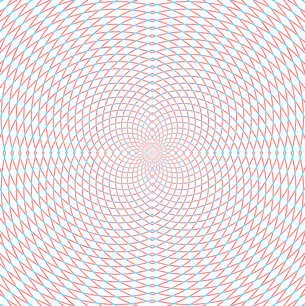
Scintillating Starburst Optical Illusion
Comprised of concentric star polygons that induce illusory ray patterns,
"scintillating starbursts" are a newly studied type of optical illusion
that highlights the subjective, constructive nature of visual
perception. Originally created as a logo for his design company, Recursia Studios,
the artist behind scintillating starbursts then teamed up with a
psychology researcher and data scientist to conduct a study on the
mechanisms underlying the effect. Their study involved 122 participants
who viewed 162 different versions of the scintillating starburst varying
in shape, complexity, and brightness. The results showed that factors
such as contrast, line width, and number of vertices influenced how
strongly the rays were perceived. The authors argue that scintillating
starbursts are a new optical illusion, distinct from grid illusions such
as the scintillating grid, Hermann grid, Motokawa grid, and pincushion grid
illusions. One of the authors explains, "[T]he rays are stronger and
more striking and at the same time fleeting or scintillating, traversing
the background." They suggest instead that scintillating starburst is a
compound illusion, combining the perception of short Motokowa line
segments that serve as guideposts for the illusory rays with a
scintillating effect from saccadic eye movements that alternate between
peripheral and foveal vision, with the effect being more pronounced to
peripheral vision. The effect is further enhanced when rotating. The
researchers also note that the illusion is an effect of luminance rather
than of chromaticity (color). One of the authors explains, "If the
background is brighter than the shape, the illusory rays will
appear brighter than the background. If the background is darker than
the shape, the illusory rays will appear darker than the background. If
background and shape are isoluminant [regardless of color], no rays
appear." While a technical study of optical illusions might seem
extraneous, it reveals the subjective and constructive nature of visual
perception. The existence and effectiveness of optical illusions
challenges the belief that what we see always corresponds with reality.
One of the authors states that a future direction could be studying the
propensity to see optical illusions with personality characteristics.
A Synthetic Norrin/Wnt Antibody Restores Blood-Retina Barrier in Diabetic Retinopathy
A team of researchers at University of Toronto are developing a new antibody as a potential treatment for diabetic retinopathy and other
diseases caused by defects in the blood-retina barrier. Specifically,
the researchers studied the Norrin/Wnt cell signalling pathway, which is
crucial for the formation and maintenance of the blood-retina barrier.
When this signalling pathway is disrupted, such as in diabetic
retinopathy, the blood vessels become leaky, compromising the
blood-retina barrier. The current publication describes how a synthetic
antibody (F4L5.13) activating the Frizzled4-LRP5 receptor complex
successfully stimulated
Norrin/Wnt signalling to restore
barrier function. This antibody activates the Norrin/Wnt pathway by
attaching to two key cell surface receptors (Frizzled4 and LRP5) and
bringing them into close proximity. The researchers have thus far tested
their antibody in cell cultures and in two mouse models, one
representing a genetic eye condition (for Norrie disease) and one for
diabetic retinopathy. In the cell culture model, the antibody restored
barrier function "in part by promoting surface expression of junction
proteins." Treatment of the antibody in mice restored barrier function
and "significantly normalized neovascularization in an oxygen-induced
retinopathy." The investigators have founded a start-up company to
proceed with translation of their work to clinical studies. One century
ago in 1921, researchers at University of Toronto developed insulin, the
life-saving medication to treat diabetes. It is especially fitting,
therefore, that U of T researchers continue that legacy, in this case to
develop a drug therapy for a common ocular complication of diabetes.
Because of similarities between the blood-retina and blood-brain
barriers, they also plan to extend their research to other contexts.
Metabolic Responses to LED and OLED Light at Night
Exposure to blue light has various effects on human health, such as in
the regulation of circadian rhythm. Researchers in Japan wanted to explore the effect of blue light on physiological processes such
as metabolism during sleep. In particular, they compared polychromatic
white LEDs (light-emitting diodes) and polychromatic white OLEDs
(organic light-emitting diodes). LEDs have been widely adopted for their
energy-saving advantages. However, polychromatic white LEDs also emit
more blue light compared to polychromatic white OLEDs, sparking
curiosity as to their potential effects on health. The study involved
ten male participants exposed to LED, OLED (1000 lux), or dim
(< 10 lux) light for 4 hours before sleep in a metabolic chamber. The
researchers then measured energy expenditure, core body temperature,
fat oxidation, and 6-sulfatoxymelatonin (melatonin) levels during
sleep. The study's senior author reports, "Although no effect on sleep
architecture was observed, energy
expenditure and core body temperature during sleep were significantly
decreased after OLED exposure. Furthermore, fat oxidation during sleep
was significantly lower after exposure to LED compared with OLED."
The results also showed a weak but significant correlation between
6-sulfatoxymelatonin levels and fat oxidation under OLED lighting. The
sample size of ten male participants is extremely small and homogeneous.
Nonetheless, given that some differences in metabolic responses were
observed among different artificial lighting conditions, connections
between artificial lighting and metabolism could be further explored in larger studies.
Illusory Faces Elicit Similar Responses as Real Faces
Face pareidolia, the perception of illusory faces in inanimate objects, lies at the intersection between optical illusions and facial recognition.
Although technically an error in visual processing, the ability to
quickly perceive faces with a crude template of two eyes, a nose, and a
mouth—and the emotions they express—is an evolutionary
benefit for judging friend or foe, a side effect that is
so common that we simply accept it as normal. Neuroscientists in Australia wanted to study the psychology behind the phenomenon,
especially as it pertains to the perception of emotions. “We know these
objects are not truly faces, yet the perception of a face
lingers,” the first author of the study explained, “We end up with
something strange: a
parallel experience that it is both a compelling face and an object. Two
things at once. The first impression of a face does not give way to the
second perception of an object.” Their study asked 17 participants to
view sequences of dozens of real and illusory faces and rate the
perceived emotion on a scale from happy to angry. The results showed
that perception of facial expression was consistent across observers,
despite variations in visual features. Secondly, the expression on prior
faces influenced the perception of subsequent faces. For example,
seeing happy faces caused the viewer to more likely perceive the next
face as happier. Lastly, this bias was observed even when the real faces
and illusory faces were interleaved in the sequence. Such a
"cross-over" condition revealed that rather than being discarded as
false detections, analysis of facial expression applied to the illusory
faces in the same way as for real faces. The lead researcher concluded,
“When objects look compellingly face-like, it is more than an
interpretation: they really are driving your brain’s face detection
network. And that scowl, or smile; that’s your brain’s facial expression
system at work. For the brain, fake or real, faces are all processed
the same way.”
In Other News
(1) Art: Optical illusion with a message about conservation
(2) Research project aims to use fluorescein video-angiogram to measure diabetic retinopathy
(3) Patients enrolled in clinical trials for presbyopia-correcting contact lenses
Sunday, August 15, 2021
Week in Review: Number 26
Subscribe to:
Post Comments (Atom)
No comments:
Post a Comment